NIR-II fluorescence lymphatic imaging and intraoperative navigation based on the “isolated cage” monodisperse strategy
摘要
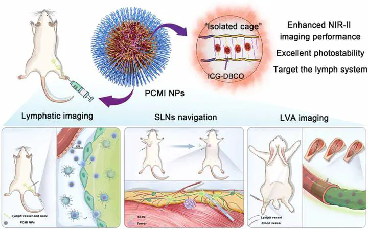
Fluorescence imaging in the second near-infrared (NIR-II) window is well suited for biological systems due to lower tissue scattering and micrometer-scale resolution at depths of millimeters. Unfortunately, none of the NIR-II fluorophores have clinical application due to low quantum yields, poor photostability, and longterm biosafety concerns. Herein, we develop NIR-II fluorescent nanoprobes based on the isolated cage monodisperse strategy, which significantly inhibits the π-π stacking interactions and restricts internal rotation of the arrested indocyanine green (ICG) fluorophores, which minimizes aggregation-caused quenching and enhances their NIR-II imaging performance. Unlike ICG’s imaging window of several minutes, the nanoformulations have reduced photobleaching and provide longer observation times. The increased imaging time allows high spatiotemporal resolution of blood vessels and lymphatic system. Benefiting from the high resolution and sensitivity of the nanoprobes, sentinel lymph nodes are accurately removed under fluorescence navigation. More intriguingly, the patency of lymphovenous anastomosis (LVA) is presented more intuitively by qualitative and quantitative NIR-II fluorescence signals, which will directly benefit the innovation and promotion of LVA.